GIANI is a FIJI plugin designed to facilitate routine analysis of 3D cell biology images. Implemented specifically with batch-processing in mind, GIANI is designed to:
- Read data sets in their native, proprietary format
- Automatically segment nuclei and cells
- Measure nuclear and cell morphology, together with fluorescence intensities within these regions.
- Allow users to explore their results
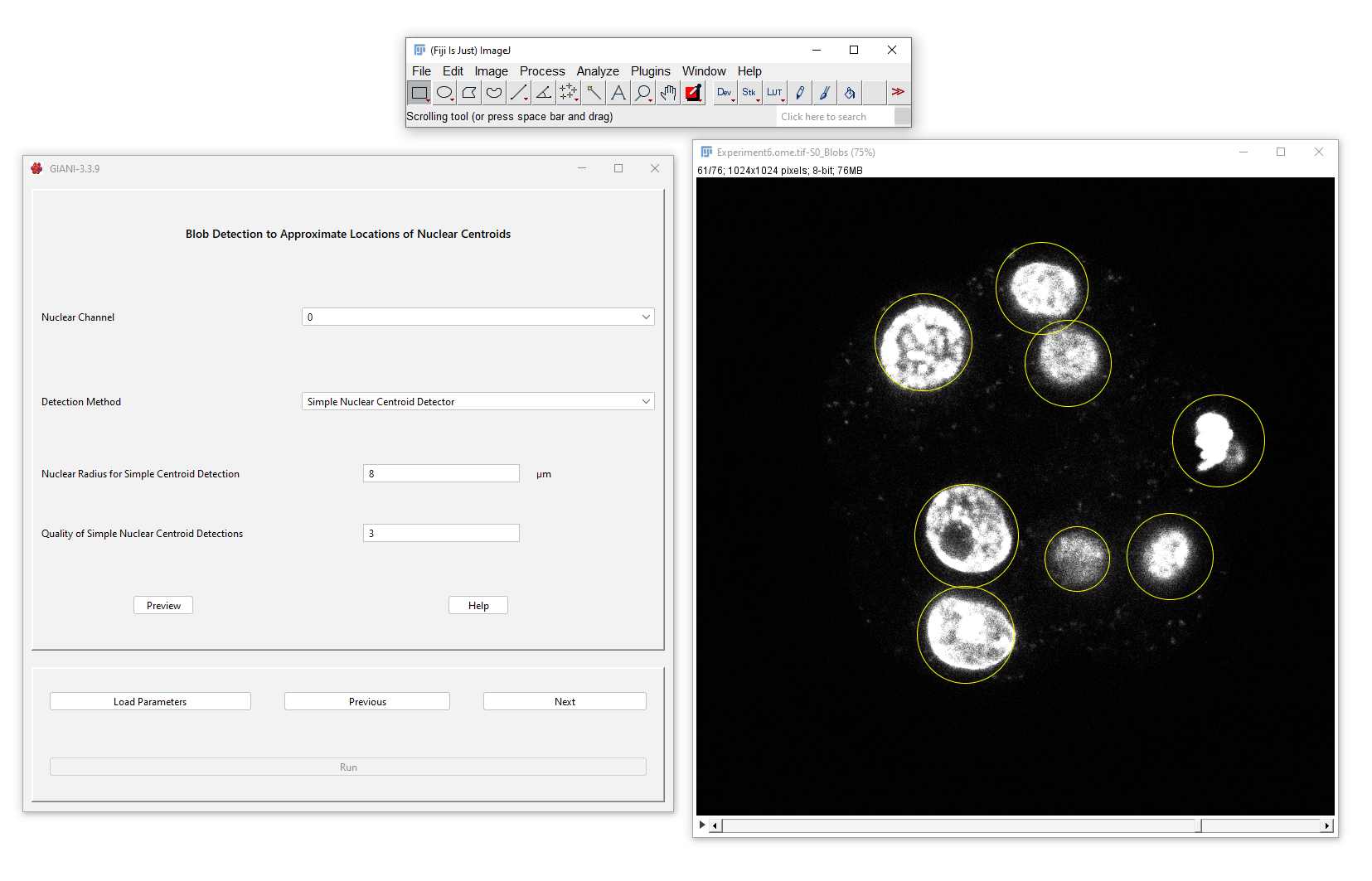
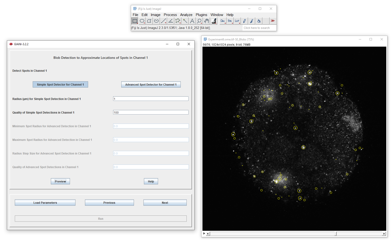
Nuclei locations are estimated using blob detection...
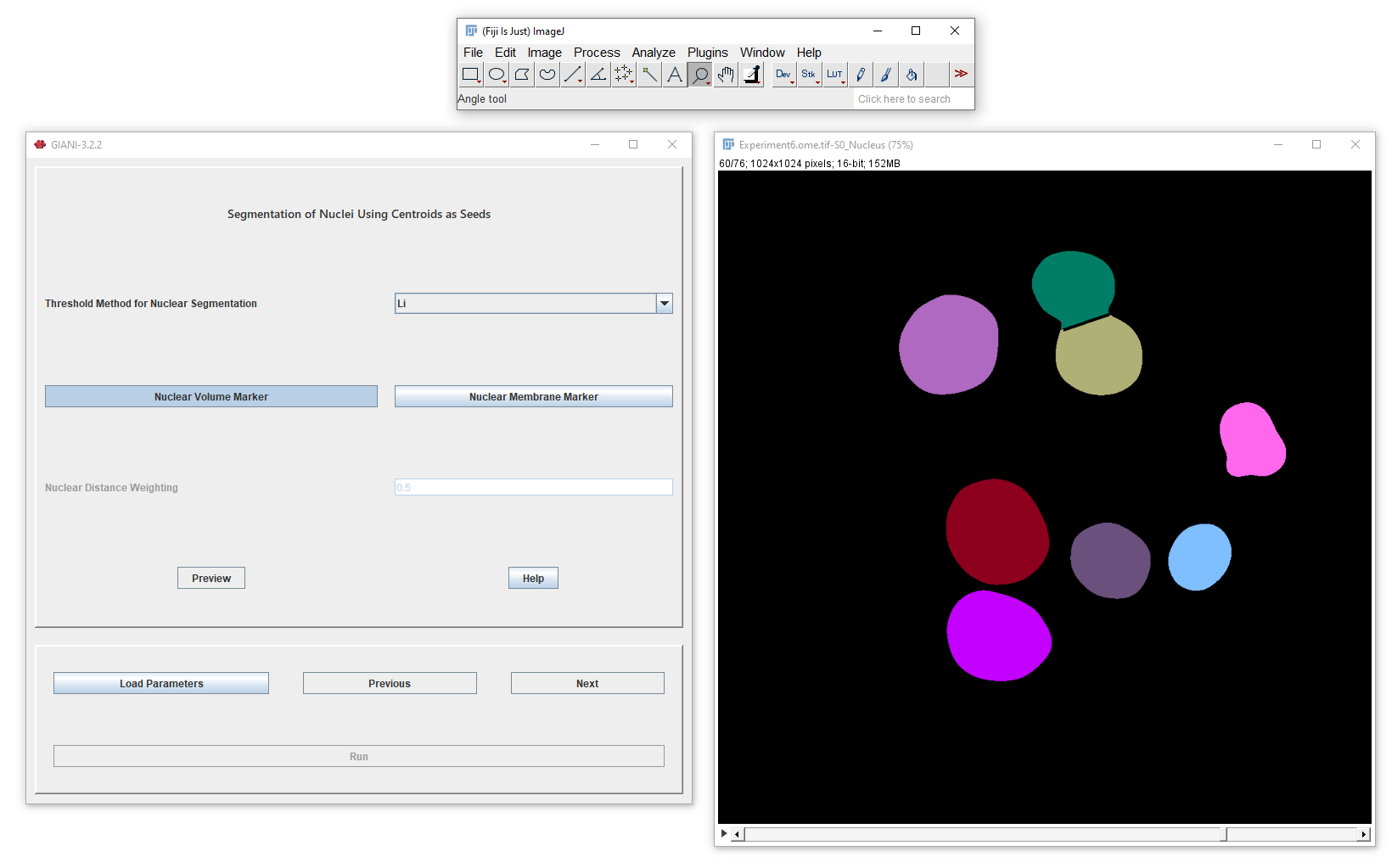
...before full segmentation is achieved using a watershed operation...
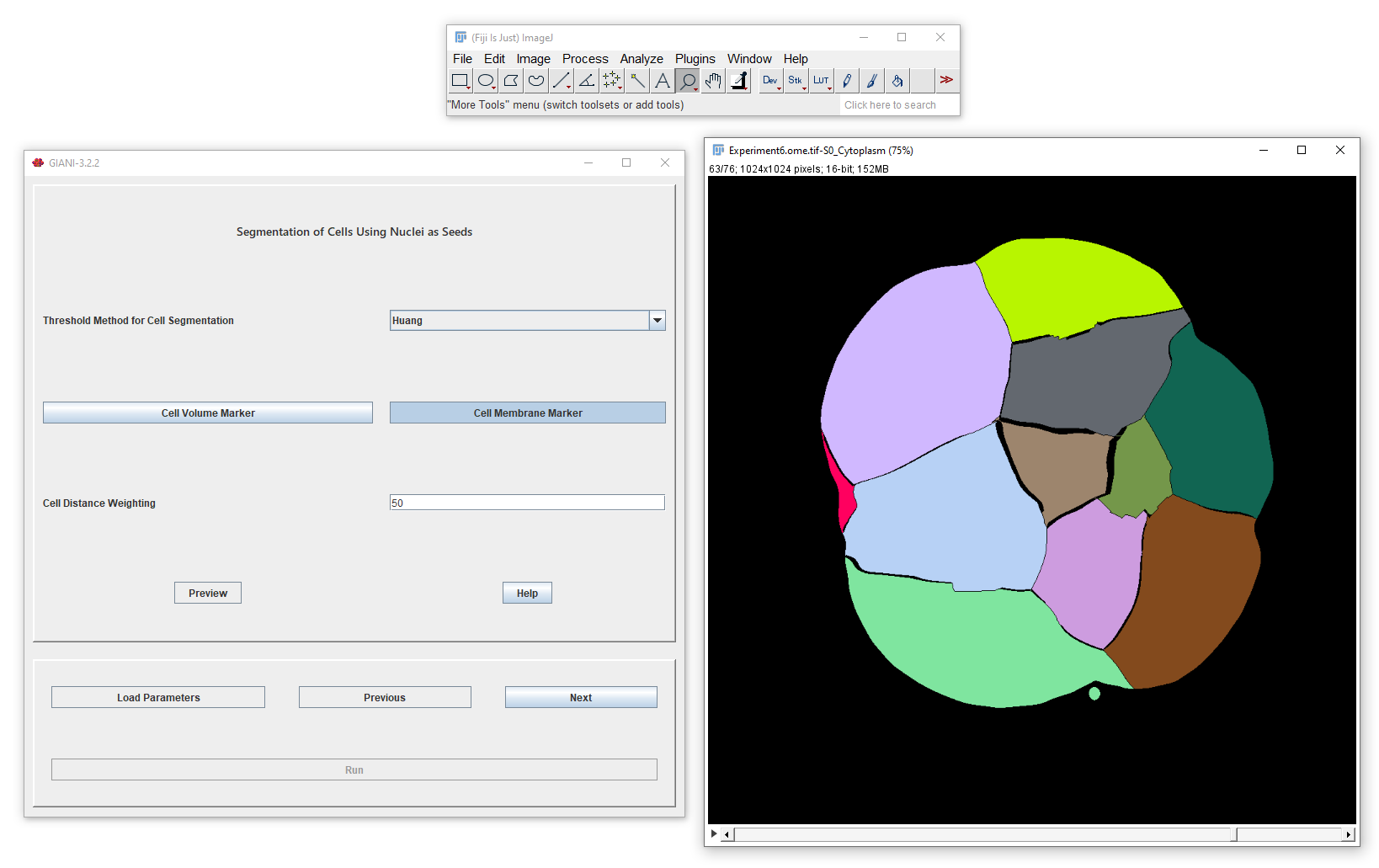
...and then cells are similarly segmented:
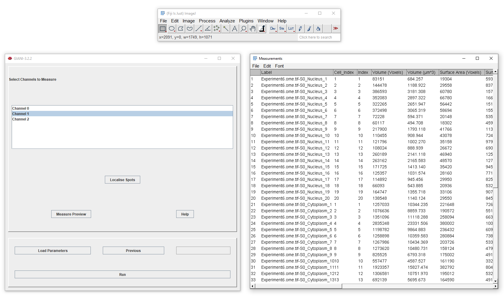
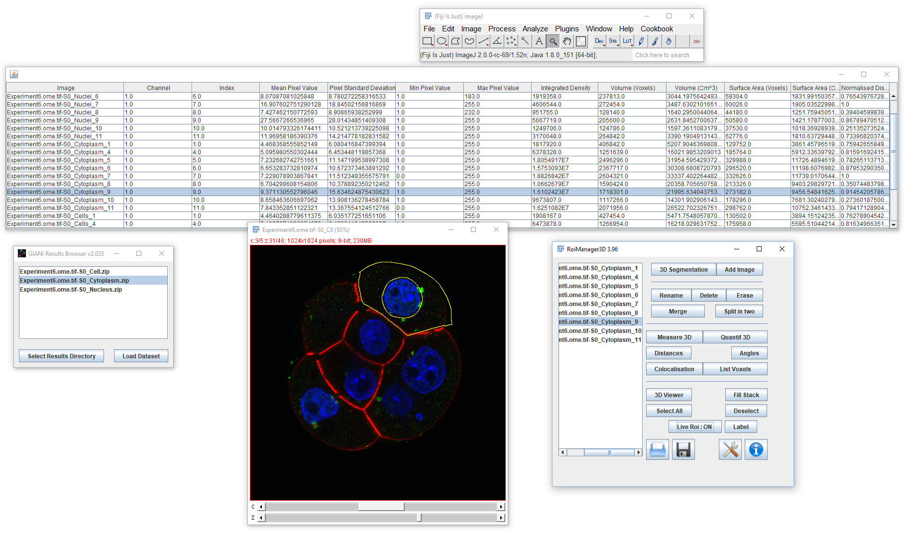
A complete set of morphological and intensity descriptors, across an unlimited number of channels, can then be obtained from the above segmentations.
If you wish, you can segment smaller, sub-cellular structures using additional blob detection:
Examine your results using the included results browser:
See the Wiki for installation instructions and full documentation.
Please cite the following publication if you use GIANI in your work:
David J. Barry, Claudia Gerri, Donald M. Bell, Rocco D'Antuono, Kathy K. Niakan; GIANI – open-source software for automated analysis of 3D microscopy images. J Cell Sci 15 May 2022; 135 (10): jcs259511. doi: https://doi.org/10.1242/jcs.259511


